Research Infrastructure at INFLPR:
– Femtosecond lasers
– Pulsed Laser Deposition systems
– Laser pyrolysis
| CETAL-PW Laser Infrastructure – 1 PW Laser System Manufacturer: Thales Optonics, France. Key features of the laser: • Peak power outputs: 1 PW / 45 TW • Ultra-short pulses, aproximately 25 fs • Spectral broadening by using XPW filter in the Front End • Pulse shaping by spectral phase and amplitude manipulation with an Acousto Optic Programmable Dispersion Filter (AOPDF) DAZZLER • High intensity ps contrast, > 1010 @100 ps • Improvement of picosecond pre pulse intensity contrast by use of XPW filter • High Strehl Ratio, more than 0.75. • Wavefront control using adaptive opticsPW – Laser  | |
| TEWALAS facility – 20 TW laser system At the beginning of the year 2009 a high power Ti:Sapphire laser was installed in our laboratory. The amplified system Pulsar20 (Amplitude Technologies) provide 10 Hz pulses with 22 fs pulse duration and max. 450 mJ pulse energy. The system is used for high harmonic generation, X-ray laser experiments, plasma experiments, high power pulsed lasers development and experiments. The system is able to output beams from the oscillator (80MHz) and from each of the amplification modules (10 Hz) at different energy level.TEWALAS laser 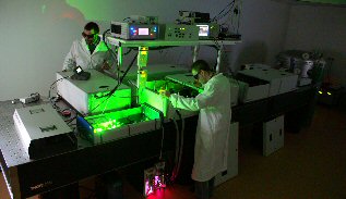 | |
| CPA – laser system Experiments with a femtosecond laser at microJules energy level and 2KHz repetition rate are done with a CPA system (Clark-MRX 2101). The amplified output beam is at 775 nm wavelength, with 200 fs pulse duration and the max. pulse energy is 0.7 mJ. This system is used in experiments such as laser ablation for micro and nano-structuring experiment on ceramics, semiconductors and metallic film, or two-photon photo-polymerization of photoresists for 3D micro-structuring, development of a multipulses laser for pump-probe experiments. An additional output from the oscillator of the CPA offers femtosecond laser beam at 35 MHz for ultrafast laser spectroscopy.CPA femtosecond laser  | |
| Nano-second and pico-second Solid-State Lasers Our group has a large experience in design and construction of flash and diode pumped solid-state lasers, laser microchip based microlasers, amplified solid-state lasers. Our most recent realisation (project NANOLAS) is a compact Nd:YAG laser with diode pumped microlaser, amplified in a multipass configuration up to 20 mJ pulse energy at 1-10 Hz. The laser can provide output beams at 1064, 532, and 266 nm, with pulse duration of about 450 ps. These laser radiations are suitable to applications such as laser microprocessing, laser induced break-down spectroscopy (LIBS), Near-field nanopatterning (see figure below)Laser near-filed nanolithography on monodispersed colloidal nanoparticles deposited on gold thin film. Laser wavelength l=532 nm. Pulse duration t=450 ps.  | |
| Microscope for Laser Direct Writing During the recent research projects we developed a microscope for Laser Direct Writing. Coupled with our femtosecond lasers, the microscope is able to produce by laser ablation any computer designed structures with submicrometer resolution on various materials. Laser induced forward transfer (LIFT) is an other method to direct write microstructures using semiconductors, metallic films, or even biological tissues. We successfully tested the two-photon photopolymerization (TPP) technique on photoresist using two different high repetition rate femtosecond lasers (the attenuated CPA laser at 2 KHz, and a femtosecond oscillator at 80 MHz). The software of our microprocessing microscope is able to create predefined 3D geometries or to import 3D files in STL format, as in the well known rapid prototyping technique.3D micro-structures on photo-polymers. 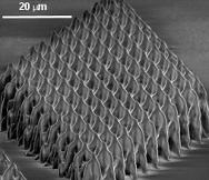 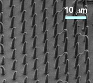 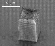 |
- Caracterizări morfologice, structurale şi compoziţionale
- Tomografie
- Laborator de biologie
- Alte echipamente
Caracterizări morfologice, structurale şi compoziţionale
High-Resolution Scanning Electron Microscopy – HRSEM
All-round nanometer or sub-nanometer resolution performance on materials ranging from nanoparticles, powders, catalysts and nanodevices to bulk magnetic samples thanks to its innovative final lens design that does not compromise on magnetic sample imaging performance.
• Compound final lens technology to boost resolution performance, providing a resolution of 1.0 nm at 1 kV without additional beam deceleration, while offering unique options for signal filtering.
• The widest range of strategies to deal with insulating samples, including: high vacuum techniques such as the SmartSCAN™ technique, drift compensated frame integration (DCFI) and charge filtering.
• Highly advanced charge mitigation strategies to deal with the most challenging applications, it is available by included low vacuum (up to 500 Pa) to mitigate charge on any sample while providing excellent resolution and large analytical currents with field-proven throughthe-lens differential pumping and dedicated LoVac detectors.
• The chamber includes a complete analytical system EDS/EBSD/WDS, an RGB-Pancromatic Cathodoluminescence module (SEM-CL), and a Kleindiek Prober-Shutle MM-PS4LTXYZ nanoprobing system.
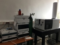
HPLC
The Agilent 6530 Q-TOF LC/MS is a liquid chromatograph-quadrupole time-of-flight mass spectrometer that performs MS/MS using a quadrupole, a hexapole collision cell and a time-of-flight analyzer to produce spectra. The system provides superior data quality and advanced analytical capabilities for profiling, identifying, characterizing, and quantifying low molecular-weight compounds and biomolecules with confidence.
The system can be used in the following application areas:
• Combinatorial chemistry target compound analysis
• Natural products and synthesized compounds screening
• Compound profiling (such as bioavailability and pK)
• Protein/peptide identification and characterization
• Metabolomics
• Biomarker discovery
• Qualitative and quantitative identification of various compounds
• Separation of compounds from mixtures
• Identifying impurities from samples (liquid or solid)
• Studying the stability of samples by identifying and characterising the degradation of compounds in time
• Determine water quality or potential food contamination
• Detection of drugs from body fluids and profiling them
• Stability determination of various compounds
• Drug metabolism
• Pharmacokinetics
• Emission and absorbtion behaviour of various compounds
• Estimation of the toxicity
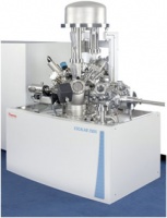
X-ray Photoelectron Spectrometer (XPS) Microprobe
Equipped with a micro-focusing X-ray monochromator designed to deliver optimum XPS performance, the ESCALAB XI+ X-ray Photoelectron Spectrometer (XPS) Microprobe ensures maximum sample throughput. The multi-technique capability and availability of a range of preparation chambers and devices provides the solution to any surface analytical problem. Using the advanced Avantage data system for acquisition and data processing, maximum information is extracted from the data.
Features:
– High sensitivity spectroscopy
– Small area XPS
– Depth profiling capability
– Angle resolved XPS.
X-ray Monochromator – Twin-crystal, micro-focusing monochromator has a 500mm Rowland circle and uses an Al anode. Sample X-ray spot size is selectable over a range of 200 to 900μm.
Lens, Analyzer and Detector – Lens/analyzer/detector combination makes the ESCALAB XI+ XPS Spectrometer unique for both imaging and small area XPS. Two types of detectors ensure optimum detection for each type of analysis — two-dimensional detector for imaging and a detector based on channel electron multipliers for spectroscopy when high count rates are to be detected. Lens is equipped with two, computer-controlled iris mechanisms — one allows the user to control the field of view of the lens down to Depth Profiling – Digitally-controlled EX06 ion gun is a high-performance ion source even when using low energy ion. Azimuthal sample rotation is available,
Electron gun can be operated at up to 1000V and provides an excellent source for REELS.
Technique Options:
– XPS with non-monochromatic X-rays
– AES (Auger electron spectroscopy)
– UPS (Ultra-violet photoelectron spectroscopy).
Tomografie
A mobile in-house built modular X-ray system used primarily as a gantry cone-beam tomograph for biology (small animals, plant roots, seeds etc.) and process tomography applications (manufacturing, environmental research). It features two modules, a gantry frame for performing gantry tomography and a frame that can accommodate the equipment for performing regular cone-beam micro-tomography (3D-CT) and X ray microbeam-fluorescence (µXRF). This multifunctional X-ray system can be used for noninvasive 3-D morphology and composition mapping.
The X-ray sources are of sealed microfocus type with maximum high voltage of 60 kV, maximum power of 50 W and different anode materials (W, Mo or Ag). X ray detection is achieved using a compact high resolution flat pannel (1944 x 1536 pixels, 75 μm pixel size) for tomography and a miniaturized X-ray spectrometer for XRF experiments. A micrometric rotation stage is used for spinning the gantry frame and four micrometric motorized stages aid in sample positioning when using the 3D-CT & µXRF module.
Customized software was developed using LabView for axis manipulation, data acquisition and control. 3-D tomographic reconstructions are obtained by a proprietary highly optimized computer code based on a modified Feldkamp algorithm.
Gantry tomography is used for samples than cannot be moved and need to lie on a sample bed rather than stand in a sample holder. It is the case of small animals, germinating seeds or samples that undergo different processes that can be analised insitu. For the inspection of miniaturised samples the microtomography analysis is guaranteed for feature recognition down to a few tens of microns. The fluorescence component can provide local qualitative and quantitative information about the sample composition elements. Using the X-Y linear stages, the µXRF system provides high resolution (~20 µm) composition mapping and accurate thickness measurements of multilayer samples.
https://erris.gov.ro/Plasma-Physics-and-Nuclear-F

Laborator de biologie
- Vertical Laminar airflow hood
Class: II biohazards; Filter: HEPA; Airflow speed: 0.4m/sec; Decontamination: UV light - Leica DM4000 B LED -Fluorescence Microscope
Working mode: transmission or reflection; Fluorescence filter: DAPI (350/50), FITC (450/490), Texas Red (562/40); Objective: 5x, 10x, 20x, 40x, 100x - Leica DMi1- Light microscope
Optical system: Infinity corrected (HCS); Contrast methods: Transmitted light: Brightfield, Phase Contrast for cell cultures. - Freezer TSE series, model TSE240GP
Manufacturer: Thermo Scientific; Name: TSE series, model TSE240GP; Capacity: 368L, 240 x2” cryobox; Dimensions: 130.8×58.7×49.3 cm HxWxD - Cell Culture CO2 incubator, Model CCl-240T-8 CO2 incubator
Volume: 170 liters; Sensor: Infrared - 3-18KS Centrifuge
Temperature Control: 4-40°C; Rotor with 4 cups; Cups adaptors d=85mm; Adaptors for 15 and 50 mL falcons - Apollo LB913 UV-VIS spectrometer
Detector: photodiode; Excitation: LED, range 400-700nm; Plate reader 96 wells; Scan read: 20s/wavelength; Shacking with 4 different amplitudes; Filters: 405nm, 450nm, 492nm and 595nm - Autoclave 8L
Volume: 8 litters; Temperature control: 20 to 130°C - CRP-18X Microplate shaker
Motor type: BLDC; Type of movement: orbital shaker 2.00mm motion; Speed: 300rpm to 1800rpm; Maximum volume: 384x60uL; Dimensions: 127x85x86 mm - CRV-45X Vortex
Type of movement: Orbital; Shaker diameter: 4.5mm; Permissible shaking weight: 100g; Maximum speed: 4500rpm

Alte echipamente
CETAL
https://cetal.inflpr.ro/newsite/equipment
http://www.erris.ro/cetal
Laboratory of Solid-State Quantum Electronics
https://erris.gov.ro/ecs-laboratory-inflpr.ro
Low Temperature Plasma Physics Department
https://erris.gov.ro/Low-Temp-Plasma-Physics
Plasma Physics and Nuclear Fusion Department
https://erris.gov.ro/Plasma-Physics-and-Nuclear-F
Lasers Departament
https://erris.gov.ro/Lasers-Department
Electron accelerators ALID and ALIN
https://erris.gov.ro/-2
Morphological, structural and compositional characterizations platform
https://erris.gov.ro/MSCCP